Paraventricular nucleus: Difference between revisions
imported>Gareth Leng |
mNo edit summary |
||
| (2 intermediate revisions by 2 users not shown) | |||
| Line 1: | Line 1: | ||
{{subpages}} | {{subpages}} | ||
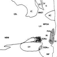
[[Image:PVNss.jpg|thumb|200 px|Human paraventricular nucleus (PVN) in this coronal section is indicated by the shaded area. Dots represent [[vasopressin]] (AVP) | [[Image:PVNss.jpg|thumb|200 px|Human paraventricular nucleus (PVN) in this coronal section is indicated by the shaded area. Dots represent [[vasopressin]] (AVP) neurones (also seen in the [[supraoptic nucleus]], SON). The medial surface is the 3rd ventricle (3V), with more lateral to the left.]] | ||
The '''paraventricular nucleus''' (PVN, PVH) is an aggregation of neurons in the [[hypothalamus]], adjacent to the third ventricle. Although it is in the periventricular zone, it is not to be confused with the [[periventricular nucleus]] | The '''paraventricular nucleus''' (PVN, PVH) is an aggregation of neurons in the [[hypothalamus]], adjacent to the third ventricle. Although it is in the periventricular zone, it is not to be confused with the nearby [[periventricular nucleus]]. The PVN is highly vascularised, but is inside the [[blood-brain barrier]], although the [[Neuroendocrinology | neuroendocrine]] neurons in this nucleus project to sites (the [[median eminence]] and the [[posterior pituitary]]) that lack a blood-brain barrier. The PVN contains [[magnocellular neurosecretory cell]]s whose axons extend into the [[posterior pituitary]], parvocellular neurosecretory cells that project to the median eminence, and several populations of [[peptide]]-containing cells that project to many different brain regions. | ||
==Magnocellular neurosecretory neurones in the PVN== | ==Magnocellular neurosecretory neurones in the PVN== | ||
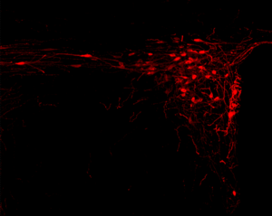
[[Image:PVN-OT.png|thumb|300 px| Oxytocin neurones immunolabelled (red) in the rat paraventricular nucleus.]] | [[Image:PVN-OT.png|thumb|300 px| Oxytocin neurones immunolabelled (red) in the rat paraventricular nucleus.]] | ||
The magnocellular cells in the PVN produce [[oxytocin]] and [[vasopressin]]. These peptide [[hormones]] are packaged in large dense-core vesicles, which are transported down the | The magnocellular cells in the PVN produce [[oxytocin]] and [[vasopressin]]. These peptide [[hormones]] are packaged in large dense-core vesicles, which are transported down the axons and released from neurosecretory nerve terminals in the [[posterior pituitary gland]]. Similar magnocellular neurones are found in the [[supraoptic nucleus]]. | ||
==Parvocellular neurosecretory neurones== | ==Parvocellular neurosecretory neurones== | ||
| Line 32: | Line 32: | ||
==References== | ==References== | ||
<references/> | <references/>[[Category:Suggestion Bot Tag]] | ||
Latest revision as of 12:01, 1 October 2024

The paraventricular nucleus (PVN, PVH) is an aggregation of neurons in the hypothalamus, adjacent to the third ventricle. Although it is in the periventricular zone, it is not to be confused with the nearby periventricular nucleus. The PVN is highly vascularised, but is inside the blood-brain barrier, although the neuroendocrine neurons in this nucleus project to sites (the median eminence and the posterior pituitary) that lack a blood-brain barrier. The PVN contains magnocellular neurosecretory cells whose axons extend into the posterior pituitary, parvocellular neurosecretory cells that project to the median eminence, and several populations of peptide-containing cells that project to many different brain regions.
Magnocellular neurosecretory neurones in the PVN
The magnocellular cells in the PVN produce oxytocin and vasopressin. These peptide hormones are packaged in large dense-core vesicles, which are transported down the axons and released from neurosecretory nerve terminals in the posterior pituitary gland. Similar magnocellular neurones are found in the supraoptic nucleus.
Parvocellular neurosecretory neurones
The parvocellular neurosecretory neurones of the PVN project axons to the median eminence, at the base of the brain. At the median eminence, the neurosecretory nerve terminals release peptides into the blood vessels of the hypothalamo-pituitary portal system. These vessels carry the peptides to the anterior pituitary gland, where they regulate hormone secretion into the systemic circulation. The parvocellular neurosecretory cells include:
- neuroendocrine cells that make corticotrophin releasing hormone (CRH), which regulates adrenocorticotropic hormone (ACTH) secretion from the anterior pituitary gland,
- neuroendocrine cells that make vasopressin: vasopressin released from these neurones also regulates ACTH secretion; vasopressin and CRH act synergistically to stimulate ACTH secretion.
- neuroendocrine cells that make thyrotrophin releasing hormone (TRH), which regulates TSH secretion.
Centrally-projecting neurons
As well as neuroendocrine neurons, the PVN contains interneurons and populations of neurons that project centrally (i.e. to other brain regions). The centrally-projecting neurons include:
- parvocellular oxytocin cells that project mainly to the brainstem and spinal cord. The oxytocin cells that project to the brainstem are involved in gastric reflexes, those that project to the spinal cord are involved in penile erection.
- parvocellular vasopressin cells that project to many areas of the hypothalamus and limbic system, as well as to the brainstem and spinal cord. These neurons are involved in blood pressure regulation and thermoregulation.
- parvocellular CRH neurons that are thought to be involved in stress-associated behaviors
The main autonomic parvocellular subcompartments of the PVN are the dorsal parvicellular PVN (dpPVN) and the ventral part of the medial parvicellular PVN (vmpPVN). The dpPVN contains mainly preganglionic sympathetic neurons projecting to the intermediolateral cell column in the spinal cord, while the vmpPVN contains most of the preganglionic parasympathetic neurons projecting to the dorsal motor nucleus of the vagus.
Afferent inputs to the PVN
The PVN receives afferent inputs from many brain regions. Amongst these, inputs from neurons in structures adjacent to the anterior wall of the third ventricle ("AV3V region") including two circumventricular organs, the subfornical organ and the organum vasculosum of the laminae terminalis, carry information about the electrolyte composition of the blood, and about circulating concentrations of hormones such as angiotensin and relaxin to regulate the magnocellular neurones. Inputs from the nucleus tractus solitarii and the ventrolateral medulla of the caudal brainstem carry information from the heart and stomach, and mediate inputs from signals received by the area postrema and afferent vagus. Inputs from the hippocampus to the CRH neurones are important regulators of stress responses. Inputs from neuropeptide Y-containing neurons in the arcuate nucleus and dorsomedial hypothalamic nucleus co-ordinate metabolic regulation via TRH secretion with regulation of energy intake, and regulate presympathetic neural activity. There is also a prominent noradrenergic innervation from the A6 cell group in the locus coeruleus. The suprachiasmatic nucleus projects strongly to the PVN to regulate circadian rhythmicity of hormone secretion.
