Central venous pressure: Difference between revisions
imported>Robert Badgett |
Pat Palmer (talk | contribs) (resolving multiple reference definition errors) |
||
| (8 intermediate revisions by 4 users not shown) | |||
| Line 1: | Line 1: | ||
{{subpages}} | {{subpages}} | ||
In physiology, the '''central venous pressure''' is "blood pressure in the central large veins of the body. It is distinguished from peripheral venous pressure which occurs in an extremity."<ref>{{MeSH}}</ref> Various disease states such as [[heart failure]] raise the central venous pressure. | {{TOC|right}} | ||
In physiology, the '''central venous pressure''' is "blood pressure in the central large veins of the body. It is distinguished from peripheral venous pressure which occurs in an extremity."<ref>{{MeSH}}</ref> Various disease states such as [[heart failure]] raise the central venous pressure. It may be approximated in physical examination, especially by [[cardiology|cardiologists]], and its continuous automated measurement is common in [[critical care]]. | |||
==Detection of elevated central venous pressure== | ==Detection of elevated central venous pressure== | ||
===Physical examination=== | ===Physical examination=== | ||
====Procedure==== | ====Procedure==== | ||
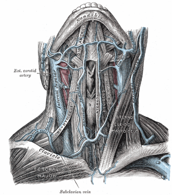
{{Image|Gray558.gif|right|350px|The veins of the neck..}} | |||
=====Inspection===== | =====Inspection===== | ||
Normal the examiner inspects the internal jugular vein while the patient sits reclined at an angle of 30° to 45°.<ref name="pmid8594245">{{cite journal |author=Cook DJ, Simel DL |title=The Rational Clinical Examination. Does this patient have abnormal central venous pressure? |journal=JAMA |volume=275 |issue=8 |pages=630–4 |year=1996 |pmid=8594245 |doi=10.1001/jama.275.8.630 |issn=}}</ref>. The jugular venous pulse is distinguished from the carotid pulse by the jugular vein showing a biphasic pulsation from 'a wave' and 'a v wave'. The jugular pulse is considered abnormal if its meniscus is 4 or more centimeters above the sternal angle of Louis. | Normal the examiner inspects the internal jugular vein while the patient sits reclined at an angle of 30° to 45°.<ref name="pmid8594245">{{cite journal |author=Cook DJ, Simel DL |title=The Rational Clinical Examination. Does this patient have abnormal central venous pressure? |journal=JAMA |volume=275 |issue=8 |pages=630–4 |year=1996 |pmid=8594245 |doi=10.1001/jama.275.8.630 |issn=}}</ref>. The jugular venous pulse (JVP) is distinguished from the carotid pulse by the jugular vein showing a biphasic pulsation from 'a wave' and 'a v wave'. The jugular pulse is considered abnormal if its meniscus is 4 or more centimeters above the sternal angle of Louis. | ||
Variations on the usual examination include using the external jugular vein<ref name="pmid4698149">{{cite journal |author=Stoelting RK |title=Evaluation of external jugular venous pressure as a reflection of right atrial pressure |journal=Anesthesiology |volume=38 |issue=3 |pages=291–4 |year=1973 |pmid=4698149 |doi= |issn=}}</ref> and examining the patient while the patient is sitting erect at 90°.<ref name="pmid18082526">{{cite journal |author=Sinisalo J, Rapola J, Rossinen J, Kupari M |title=Simplifying the estimation of jugular venous pressure |journal=Am. J. Cardiol. |volume=100 |issue=12 |pages=1779–81 |year=2007 |pmid=18082526 |doi=10.1016/j.amjcard.2007.07.030 |issn=}}</ref> When examining the patient sitting upright, the jugular pulse is considered abnormal if it is visualized above the clavicle. | Variations on the usual examination include using the external jugular vein<ref name="pmid4698149">{{cite journal |author=Stoelting RK |title=Evaluation of external jugular venous pressure as a reflection of right atrial pressure |journal=Anesthesiology |volume=38 |issue=3 |pages=291–4 |year=1973 |pmid=4698149 |doi= |issn=}}</ref> and examining the patient while the patient is sitting erect at 90°.<ref name="pmid18082526">{{cite journal |author=Sinisalo J, Rapola J, Rossinen J, Kupari M |title=Simplifying the estimation of jugular venous pressure |journal=Am. J. Cardiol. |volume=100 |issue=12 |pages=1779–81 |year=2007 |pmid=18082526 |doi=10.1016/j.amjcard.2007.07.030 |issn=}}</ref> | ||
The JVP is considered abnormal when the meniscus of the pulse is 4 cm above the sternal angle of Louis when the patient is reclining at 45°. When examining the patient sitting upright, the jugular pulse is considered abnormal if it is visualized above the clavicle. When the pulsation is elevated the patient has jugular venous distention (JVD). | |||
=====Abdominojugular test===== | =====Abdominojugular test===== | ||
| Line 15: | Line 19: | ||
====Interpretation==== | ====Interpretation==== | ||
The physical examination is more [[specificity (tests)|specific]] than [[sensitivity (tests)|sensitive]] in detecting an elevated central venous pressure according to a [[systematic review]] by the [http://www.sgim.org/clinexam-rce.cfm Rational Clinical Examination] (RCE).<ref name="pmid8594245" | |||
{| class="wikitable" | |||
|+ Accuracy of the jugular venous distention and [[abdominojugular test]].<ref name="pmid8594245"/><ref name="pmid9169900">Review: Subtle clinical findings can detect left-sided heart failure in adults. ACP J Club. 1998 Jan-Feb;128(1):11. Review of [http://pubmed.gov/9169900 PMID 9169900]</ref><ref name="pmid3415106"/><ref name="pmid2182296"/> | |||
! !!colspan="2"|Increased<br/>central venous pressure!! colspan="2"|Increased<br/>left ventricular end diastolic pressure | |||
|- | |||
! !![[Sensitivity and specificity|Sensitivity]]!![[Sensitivity and specificity|Specificity]]!![[Sensitivity and specificity|Sensitivity]]!![[Sensitivity and specificity|Specificity]] | |||
|- | |||
| Jugular venous distention|| 48%<ref name="pmid8594245"/>||88%<ref name="pmid8594245"/>|| 55% to 65%<ref name="pmid9169900"/>|| 74% to 80%<ref name="pmid9169900"/> | |||
|- | |||
| [[Abdominojugular test]]||24% to 72%<ref name="pmid3415106"/><ref name="pmid2182296"/>||96% to 93<ref name="pmid3415106"/><ref name="pmid2182296"/>|| || | |||
|} | |||
The physical examination of jugular venous distention is more [[specificity (tests)|specific]] than [[sensitivity (tests)|sensitive]] in detecting an elevated central venous pressure according to a [[systematic review]] by the [http://www.sgim.org/clinexam-rce.cfm Rational Clinical Examination] (RCE).<ref name="pmid8594245"/> The accuracy of visualizing the meniscus of the jugular pulse at 4 or more centimers above the sternal angle of Louis in the reclining patient according to one study in included in the [[systematic review]] by the RCE:<ref name="pmid2316561">{{cite journal |author=Cook DJ |title=Clinical assessment of central venous pressure in the critically ill |journal=Am. J. Med. Sci. |volume=299 |issue=3 |pages=175–8 |year=1990 |pmid=2316561 |doi= |issn=}}</ref> | |||
* [[sensitivity (tests)|sensitivity]] = 48% | * [[sensitivity (tests)|sensitivity]] = 48% | ||
* [[specificity (tests)|specificity]] = 88% | * [[specificity (tests)|specificity]] = 88% | ||
| Line 22: | Line 39: | ||
* [[sensitivity (tests)|sensitivity]] = 77% | * [[sensitivity (tests)|sensitivity]] = 77% | ||
* [[specificity (tests)|specificity]] = 68% | * [[specificity (tests)|specificity]] = 68% | ||
An elevated JVD is associated with reduced prognosis among patients with [[heart failure]].<ref name="pmid11529211">{{cite journal| author=Drazner MH, Rame JE, Stevenson LW, Dries DL| title=Prognostic importance of elevated jugular venous pressure and a third heart sound in patients with heart failure. | journal=N Engl J Med | year= 2001 | volume= 345 | issue= 8 | pages= 574-81 | pmid=11529211 | |||
| url=http://www.ncbi.nlm.nih.gov/entrez/eutils/elink.fcgi?dbfrom=pubmed&tool=clinical.uthscsa.edu/cite&email=badgett@uthscdsa.edu&retmode=ref&cmd=prlinks&id=11529211 }}</ref> | |||
In a patient with [[shock (physiology)|shock]] central venous pressure of >7 cm H<sub>2</sub>O suggest [[cardiogenic shock]].<ref name="pmid20945471">{{cite journal| author=Vazquez R, Gheorghe C, Kaufman D, Manthous CA| title=Accuracy of bedside physical examination in distinguishing categories of shock: a pilot study. | journal=J Hosp Med | year= 2010 | volume= 5 | issue= 8 | pages= 471-4 | pmid=20945471 | doi=10.1002/jhm.695 | pmc= | url= }} </ref> | |||
==References== | ==References== | ||
<references/> | <small> | ||
<references> | |||
</references> | |||
</small> | |||
[[Category:Suggestion Bot Tag]] | |||
Latest revision as of 14:42, 19 September 2024
In physiology, the central venous pressure is "blood pressure in the central large veins of the body. It is distinguished from peripheral venous pressure which occurs in an extremity."[1] Various disease states such as heart failure raise the central venous pressure. It may be approximated in physical examination, especially by cardiologists, and its continuous automated measurement is common in critical care.
Detection of elevated central venous pressure
Physical examination
Procedure
Inspection
Normal the examiner inspects the internal jugular vein while the patient sits reclined at an angle of 30° to 45°.[2]. The jugular venous pulse (JVP) is distinguished from the carotid pulse by the jugular vein showing a biphasic pulsation from 'a wave' and 'a v wave'. The jugular pulse is considered abnormal if its meniscus is 4 or more centimeters above the sternal angle of Louis.
Variations on the usual examination include using the external jugular vein[3] and examining the patient while the patient is sitting erect at 90°.[4]
The JVP is considered abnormal when the meniscus of the pulse is 4 cm above the sternal angle of Louis when the patient is reclining at 45°. When examining the patient sitting upright, the jugular pulse is considered abnormal if it is visualized above the clavicle. When the pulsation is elevated the patient has jugular venous distention (JVD).
Abdominojugular test
The abdominojugular test (AJR) is another method of detecting an abnormal central venous pressure. According to some[5], but not all[6] studies, the AJR is more sensitive than inspection.
Interpretation
| Increased central venous pressure |
Increased left ventricular end diastolic pressure | |||
|---|---|---|---|---|
| Sensitivity | Specificity | Sensitivity | Specificity | |
| Jugular venous distention | 48%[2] | 88%[2] | 55% to 65%[7] | 74% to 80%[7] |
| Abdominojugular test | 24% to 72%[5][6] | 96% to 93[5][6] | ||
The physical examination of jugular venous distention is more specific than sensitive in detecting an elevated central venous pressure according to a systematic review by the Rational Clinical Examination (RCE).[2] The accuracy of visualizing the meniscus of the jugular pulse at 4 or more centimers above the sternal angle of Louis in the reclining patient according to one study in included in the systematic review by the RCE:[8]
- sensitivity = 48%
- specificity = 88%
If inspecting the patient sitting erect, visualizing the jugular pulse above the clavicle is:[4]
- sensitivity = 77%
- specificity = 68%
An elevated JVD is associated with reduced prognosis among patients with heart failure.[9]
In a patient with shock central venous pressure of >7 cm H2O suggest cardiogenic shock.[10]
References
- ↑ Anonymous (2024), Central venous pressure (English). Medical Subject Headings. U.S. National Library of Medicine.
- ↑ 2.0 2.1 2.2 2.3 2.4 Cook DJ, Simel DL (1996). "The Rational Clinical Examination. Does this patient have abnormal central venous pressure?". JAMA 275 (8): 630–4. DOI:10.1001/jama.275.8.630. PMID 8594245. Research Blogging.
- ↑ Stoelting RK (1973). "Evaluation of external jugular venous pressure as a reflection of right atrial pressure". Anesthesiology 38 (3): 291–4. PMID 4698149. [e]
- ↑ 4.0 4.1 Sinisalo J, Rapola J, Rossinen J, Kupari M (2007). "Simplifying the estimation of jugular venous pressure". Am. J. Cardiol. 100 (12): 1779–81. DOI:10.1016/j.amjcard.2007.07.030. PMID 18082526. Research Blogging.
- ↑ 5.0 5.1 5.2 5.3 Ewy GA (1988). "The abdominojugular test: technique and hemodynamic correlates". Ann. Intern. Med. 109 (6): 456–60. PMID 3415106. [e]
- ↑ 6.0 6.1 6.2 6.3 Marantz PR, Kaplan MC, Alderman MH (1990). "Clinical diagnosis of congestive heart failure in patients with acute dyspnea". Chest 97 (4): 776–81. PMID 2182296. [e]
- ↑ 7.0 7.1 7.2 Review: Subtle clinical findings can detect left-sided heart failure in adults. ACP J Club. 1998 Jan-Feb;128(1):11. Review of PMID 9169900
- ↑ Cook DJ (1990). "Clinical assessment of central venous pressure in the critically ill". Am. J. Med. Sci. 299 (3): 175–8. PMID 2316561. [e]
- ↑ Drazner MH, Rame JE, Stevenson LW, Dries DL (2001). "Prognostic importance of elevated jugular venous pressure and a third heart sound in patients with heart failure.". N Engl J Med 345 (8): 574-81. PMID 11529211.
- ↑ Vazquez R, Gheorghe C, Kaufman D, Manthous CA (2010). "Accuracy of bedside physical examination in distinguishing categories of shock: a pilot study.". J Hosp Med 5 (8): 471-4. DOI:10.1002/jhm.695. PMID 20945471. Research Blogging.
