Human brain: Difference between revisions
Jump to navigation
Jump to search


imported>Daniel Mietchen (+image) |
imported>Daniel Mietchen (image legend) |
||
| Line 1: | Line 1: | ||
{{subpages}} | {{subpages}} | ||
<!-- Text is transcluded from the BASEPAGENAME/Definition subpage--> | <!-- Text is transcluded from the BASEPAGENAME/Definition subpage--> | ||
{{Image|Brain-MRI-location-guide.png|right|350px|[[MRI]] scan of a | {{Image|Brain-MRI-location-guide.png|right|350px|[[MRI]] scan of a human brain, with several key anatomical structures labeled. Abbreviations: CC, [[cerebellum]]; DMT, dorsal medial [[thalamus]]; Hypo, [[hypothalamus]]; LAI/RAI, left/right anterior [[insula]]; LAP/RAP, left/right posterior insula; LCN/RCN, left/right [[caudate nucleus]]; LG, [[lingual gyrus]]; LHipp/RHipp, left/right [[hippocampus]]; LI/RI, left/right insula; PLT, posterior lateral thalamus; VLT, ventral lateral thalamus. Distances from the [[anterior commissure]] and orientation, based on the standard [[Montreal Neurological Institute space]], are (A) 5 mm superior, (B) 5 mm left, (C) 27 mm right, and (D) 19 mm posterior. Distance increases from left-to-right for [[sagittal]] (side) views, posterior-to-anterior for [[coronal]] views, and inferior to superior for [[axial]] (transverse) views. The background image is a high-resolution scan from a single participant (normalized to Montreal Neurological Institute space).}} | ||
Revision as of 05:39, 23 May 2010
Human brain [r]: The core unit of the central nervous system in our species. [e]
This article contains just a definition and optionally other subpages (such as a list of related articles), but no metadata. Create the metadata page if you want to expand this into a full article.

(CC) Image: Macey et al., 2007
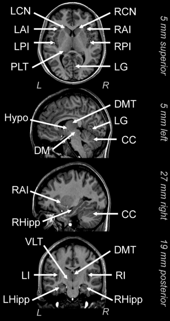
MRI scan of a human brain, with several key anatomical structures labeled. Abbreviations: CC, cerebellum; DMT, dorsal medial thalamus; Hypo, hypothalamus; LAI/RAI, left/right anterior insula; LAP/RAP, left/right posterior insula; LCN/RCN, left/right caudate nucleus; LG, lingual gyrus; LHipp/RHipp, left/right hippocampus; LI/RI, left/right insula; PLT, posterior lateral thalamus; VLT, ventral lateral thalamus. Distances from the anterior commissure and orientation, based on the standard Montreal Neurological Institute space, are (A) 5 mm superior, (B) 5 mm left, (C) 27 mm right, and (D) 19 mm posterior. Distance increases from left-to-right for sagittal (side) views, posterior-to-anterior for coronal views, and inferior to superior for axial (transverse) views. The background image is a high-resolution scan from a single participant (normalized to Montreal Neurological Institute space).
MRI scan of a human brain, with several key anatomical structures labeled. Abbreviations: CC, cerebellum; DMT, dorsal medial thalamus; Hypo, hypothalamus; LAI/RAI, left/right anterior insula; LAP/RAP, left/right posterior insula; LCN/RCN, left/right caudate nucleus; LG, lingual gyrus; LHipp/RHipp, left/right hippocampus; LI/RI, left/right insula; PLT, posterior lateral thalamus; VLT, ventral lateral thalamus. Distances from the anterior commissure and orientation, based on the standard Montreal Neurological Institute space, are (A) 5 mm superior, (B) 5 mm left, (C) 27 mm right, and (D) 19 mm posterior. Distance increases from left-to-right for sagittal (side) views, posterior-to-anterior for coronal views, and inferior to superior for axial (transverse) views. The background image is a high-resolution scan from a single participant (normalized to Montreal Neurological Institute space).