Oxytocin
Oxytocin (Greek: "quick birth") is a mammalian hormone that is secreted from the pituitary gland into the blood, but which is also released into the brain. In women, it is secreted into the blood during labor to facilitate childbirth, and when a baby is sucks at the nipple to facilitate breastfeeding. Oxytocin is also released during sexual intercourse, at orgasm, in both men and women. In the brain, oxytocin is involved in sexual behavior, regulating food intake, social recognition and bonding, and, as suggested by recent research, it might be involved in the formation of "trust" between people.
Synthesis, storage and release
Oxytocin is made by a population of neurons in the hypothalamus - specifically, by magnocellular neurosecretory cells of the supraoptic nucleus and paraventricular nucleus. These neurons are larger than most neurons in the hypothalamus; in the rat their cell bodies have a diameter of 15-20 micrometres. These neurons secrete oxytocin into the blood from nerve endings in the posterior lobe of the pituitary gland (also called the "neurohypophysis", or neural lobe). Oxytocin is also made by smaller ("parvocellular") neurons in the paraventricular nucleus that project to other parts of the brain and to the spinal cord.
In the pituitary gland, oxytocin is packaged in dense-core vesicles, where it is bound to neurophysin, as shown in the inset of the figure. Neurophysin is a peptide fragment of the precursor protein molecule from which oxytocin is derived by enzymatic cleavage; whenever oxytocin is secreted, neurophysin is also secreted, along with other fragments of the precursor, but only oxytocin is known to have physiological actions after release. Typically one oxytocin neuron has just a single axon that projects to the posterior pituitary, but this axon gives rise to about 10,000 nerve terminals, each of which may contain many thousands of vesicles. Each vesicle contains about 10,000 molecules of oxytocin.
Oxytocin secretion is controlled by the electrical activity of the oxytocin cells in the hypothalamus. These cells generate action potentials that propagate down axons to the nerve endings in the pituitary, and briefly depolarise the neurosecretory nerve terminals; whenever the nerve terminals are depolarised, some of the oxytocin-containing vesicles will be released by exocytosis.
Oxytocin and vasopressin are the only "true" hormones secreted from the human posterior pituitary gland. However, oxytocin neurons make (and secrete) several other peptides, including cholecystokinin and dynorphin; these act either within the hypothalamus or within the posterior pituitary gland, but too little of these is secreted for them to have any effect at distant targets. The magnocellular neurons that make oxytocin are mingled amongst other magnocellular neurons that make vasopressin; vasopressin neurons are in many respects, very similar to the oxytocin neurons.
Actions of Oxytocin
Oxytocin has well known peripheral (hormonal) actions, but also has actions in the brain. All of the effects of oxytocin are mediated by specific, high affinity oxytocin receptor molecules. [1] Oxytocin receptors belong to the rhodopsin-type (class I) group of G-protein-coupled receptors; theu are expressed in the myometrium and endometrium of the uterus during pregnancy, and on the myoepithelial cells of the mammary gland in lactation. In various species, oxytocin receptors are also expressed at other peripheral sites, including the kidneys, vas deferens and heart. In the brain, oxytocin receptors are expressed at several sites, but the precise distribution differs between species. Many areas of the hypothalamus are rich in oxytocin receptors, and receptors are also expressed in the caudal brainstem (especially in the nucleus of the solitary tract), the spinal cord, the hippocampus, the septum, the amygdala and the olfactory bulbs. At each of these sites oxytocin receptors are expressed by discrete subpopulations of neurons, not by all neurons in that area. Only one type of specific oxytocin receptor has been identified, and the structure of this receptor has been highly conserved through evolution.
Peripheral (hormonal) actions
The peripheral actions of oxytocin mainly reflect secretion from the pituitary gland, but there is some evidence that some oxytocin is also produced in the uterus during pregnancy in some species.
- Milk letdown In breastfeeding mothers, oxytocin acts at the mammary glands, causing milk to be 'let down' into a collecting chamber, from where it can be extracted by sucking at the nipple. When an infant sucks at the nipple, sensory receptors are activated, and this information is relayed by spinal nerves to the hypothalamus, and ultimately causes the neurons that make oxytocin to fire action potentials in intermittent bursts. These bursts result in the secretion of large pulses of oxytocin secretion from the neurosecretory nerve terminals in the pituary gland.
- Uterine contraction Oxytocin is an important hormone for cervical dilation before birth, and it causes uterine contractions during the second and third stages of labor. Oxytocin acts on the myometrium to induce contractions, but also on the endometrium to stimulate the production of prostaglandins, which also can stimulate uterine contractions. The uterine contractions in turn cause more oxytocin to be secreted from the pituitary gland, in a positive-feedback loop (the "Ferguson reflex"). Oxytocin secretion during breastfeeding can continue to cause uterine contractions during the first few weeks of lactation; these are usually mild but sometimes can be painful. The actions of oxytocin upon the uterus are part of the physiological mechanisms for expelling the fetus and placenta, and they also assist the uterus in "clotting" the placental attachment after birth. Oxytocin (or in marsupials, the closely related peptide mesotocin) seems to play a role in parturition in all mammalian species; it is secreted at high levels during labor, and uterine sensitivity to oxytocin as at its maximum just before birth begins. However, although important, oxytocin is not essential for normal parturition; in transgenic mice that cannot make any oxytocin, or mice that cannot make any receptors for oxytocin (so-called "gene knockout" mice), parturition is normal, although the mice are unable to feed their young because no milk is let down in response to suckling.[2]
- Oxytocin is secreted into the blood at orgasm in both males and females [3] In males, oxytocin may facilitate sperm transport in ejaculation.
- Because of its similarity to vasopressin, high doses of oxytocin can affect the excretion of urine slightly (oxutocin is a weak agonist at vasopressin receptors). More important, in several species, oxytocin can stimulate sodium excretion from the kidneys (natriuresis), and in humans, high doses of oxytocin can result in hyponatremia.
- Oxytocin and oxytocin receptors are found in the heart in some rodents, and the hormone may play a role in the embryonal development of the heart by promoting cardiomyocyte differentiation. [4]. However, the absence of either oxytocin or its receptor in knockout mice has not been reported to produce cardiac insufficiencies.
Actions in the brain
Oxytocin secreted into the blood cannot enter the brain because of the blood-brain barrier. Nevertheless, injections of very small amounts of oxytocin into the brain have several different effects on behavior. In particular, oxytocin facilitates sexual behaviors (in males and in females), facilitates maternal behavior and "bonding" between partners, facilitates "grooming" behavior (in rodents), reduces ingestive behaviors (inhibits salt appetite in rodents), and it seems to ameliorate or counteract some of the effects of stress on behavior. Injections of oxytocin antagonists tend to have the opposite effects to those of oxytocin, so it seems likely that these are behaviors which are normally regulated by oxytocin released within the brain.
The behavioral effects of oxytocin might reflect release either from the nerve endings of centrally-projecting oxytocin neurons, or from the dendrites of the magnocellular oxytocin neurons. The centrally-projecting oxytocin neurons are small ("parvocellular") neurons, separate from the large ("magnocellular") neurons that project (send axons) to the pituitary gland); they project mainly to the caudal brainstem and spinal cord. Oxytocin receptors are found on neurons in many parts of the brain and spinal cord, including the amygdala, ventromedial hypothalamus, septum, and the nucleus of the solitary tract in the caudal brainstem.
- Sexual arousal Oxytocin injected into the cerebrospinal fluid can cause erections in male rats, reflecting actions both in the hypothalamus and spinal cord.
- Bonding In the Prairie Vole, oxytocin released into the brain of the female during sexual activity is important for forming a monogamous pair bond with her sexual partner. (Vasopressin appears to have a similar effect in males [2]). In people, plasma concentrations of oxytocin have been reported to be higher amongst people who claim to be "falling in love". Oxytocin has a role in social behaviors in many species, and so might have similar roles in humans. It has been suggested that deficiencies in oxytocin pathways in the brain might be a feature of autism.
- Maternal behavior Ewes and female rats show maternal behavior towards their offspring shortly after giving birth. In rats, this takes the form of building a nest, and gathering the newborn pups into the nest, and crouching over them to warm them and to allow them to suckle. Rats will show this behavior to any pup that is introduced into their cage, not only to their own young. Ewes on the other hand recognise their own lambs, allowing them to suckle, but rebuff all strange lambs. Neither ewes nor rats exhibit typical maternal behavior if they are given oxytocin antagonists into the brain after giving birth. However, ewes can be induced to show maternal behavior towards foreign lambs by injections of oxytocin into the brain. Rats do not show normal maternal behavior if they have given birth, for instance, by cesarean section, but can be induced to show maternal behavior by oxytocin injections into the brain[3]
- Anti-stress functions Oxytocin injected into the brain reduces blood pressure, reduces secretion of the stress hormone ACTH, increases tolerance to pain, and reduces anxiety. Oxytocin might play a role in encouraging "tend and befriend", as opposed to "fight or flight", behavior, in response to stress.
- Increasing trust and reducing fear. In a "risky investment game", volunteers who were given given nasally administered oxytocin displayed much more "trust" than a control group. Subjects who were told that they were interacting with a computer showed no such reaction, leading to the conclusion that oxytocin was not merely affecting risk-aversion[5]. Nasally-administered oxytocin was also reported to reduce fear, perhaps by its effects on the amygdala, which is thought to be responsible for fear responses. [6]
Structure and relation to vasopressin
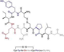
Oxytocin is a nonapeptide (i.e. it has nine amino acids), with a molecular mass of 1007 daltons. One international unit (IU) of oxytocin is equivalent to about 2 micrograms of peptide. The amino acid sequence is: cysteine-tyrosine-isoleucine-glutamine-asparagine-cysteine-proline-leucine-glycine
The cysteine residues form a sulfur bridge. This sequence is very like that of vasopressin, which is also a nonapeptide with a sulfur bridge, whose sequence differs from oxytocin by two amino acids:
cysteine-tyrosine-phenylalanine-glutamine-asparagine-cysteine-proline- arginine-glycine
Sequences of some other members of the vasopressin/oxytocin superfamily and the species expressing them are given in the vasopressin article. Oxytocin and vasopressin were discovered, isolated and synthesized by Vincent du Vigneaud in 1953, work for which he won the Nobel Prize in Chemistry in 1955.
Uses
Synthetic oxytocin is sold as medication under the names Pitocin and Syntocinon and also as generic Oxytocin. Oxytocin is destroyed in the gastrointestinal tract, and so must be administered by injection into the blood stream (for effects on peripheral organs) or by nasal spray (to reach brain areas). Oxytocin given intravenously does not enter the brain in significant quantities - it is excluded from the brain by the blood-brain barrier. Drugs administered by nasal spray are thought to have better access to the central nervous system. An oxytocin nasal spray has been used to stimulate breastfeeding; oxytocin acts in the brain to facilitate the activity of the oxytocin neurons.Oxytocin has a half-life of about three minutes in the blood.
Oxytocin analogues are used in obstetrics to induce labour and support labour in case of non-progression of parturition, and has largely replaced ergotamine as the principal agent to increase uterine tone in acute postpartum haemorrhage. Oxytocin is also used in veterinary medicine to facilitate birth and to increase milk production. The tocolytic agent atosiban (Tractocile®) is an antagonist of oxytocin receptors; it is registered in many countries to suppress premature labour between 24 and 33 weeks of gestation, and is reported to have fewer side-effects than drugs previously used for this (ritodrine, salbutamol and terbutaline).
Evolution
All vertebrates have an oxytocin-like nonapeptide hormone that supports reproductive functions and a vasopressin-like nonapeptide hormone involved in water regulation. The two genes are always located close to each other (less than 15,000 bases apart) on the same chromosome, and are transcribed in opposite directions. It is thought that the two genes resulted from a gene duplication event; the ancestral gene is estimated to be about 500 million years old and is found in cyclostomes (modern members of the Agnatha).
References
- ↑ Gimpl G, Fahrenholz F (2001) The oxytocin receptor system: structure, function, and regulation Physiol Rev 81: full text PMID 11274341
- ↑ Takayanagi Y et al (2005) Pervasive social deficits, but normal parturition, in oxytocin receptor-deficient mice. Proc Natl Acad Sci USA 102:16096-101 PMID 16249339
- ↑ Carmichael MSet al (1987) Plasma oxytocin increases in the human sexual response. J Clin Endocrinol Metab 64:27-31 PMID 3782434
- ↑ Jankowski et al (2004) Oxytocin in cardiac ontogeny. Proc Natl Acad Sci USA 101:13074-9 online PMID 15316117
- ↑ Kosfeld M et al (2005) Oxytocin increases trust in humans Nature 435:673-76 PMID 15931222
- ↑ Kirsch P et al (2005) Oxytocin modulates neural circuitry for social cognition and fear in humans J Neurosci 25:11489-93 PMID 16339042 [1]
- Caldwell HK, Young WS III. (2006) Oxytocin and vasopressin: genetics and behavioral implications. In Lim R (ed.) Handbook of Neurochemistry and Molecular Neurobiology 3rd edition, Springer, New York 320kb PDF
News stories
- Economist.com - 'Paying through the nose: A person's level of trust can be changed with a chemical spray', The Economist (June 2, 2005)
- NewScientist.com - 'Trust me, I’m spraying you with hormones' (report on trust study), Andy Coghlan, New Scientist (June 1, 2005
- NewScientist.com - 'Release of Oxytocin due to penetrative sex reduces stress and neurotic tendencies', New Scientist (January 26, 2006)
- NIH.gov - 'Oxytocin (Systemic)' (drug information), National Institute of Medicine
- Oxytocin.org - 'I get a kick out of you: Scientists are finding that, after all, love really is down to a chemical addiction between people', The Economist (February 12, 2004)
- SMH.com.au - 'To sniff at danger: Inhalable oxytocin could become a cure for social fears', Boston Globe (January 12, 2006)
oxytocin, prepro- (neurophysin I)
| |
| Identifiers | |
| Symbol(s) | OXT OT |
| Entrez | 5020 |
| OMIM | 167050 |
| RefSeq | NM_000915 |
| UniProt | P01178 |
| Other data | |
| Locus | Chr. 20 p13 |
oxytocin receptor
| |
| Identifiers | |
| Symbol(s) | OXTR |
| Entrez | 5021 |
| OMIM | 167055 |
| RefSeq | NM_000916 |
| UniProt | P30559 |
| Other data | |
| Locus | Chr. 3 p25 |
